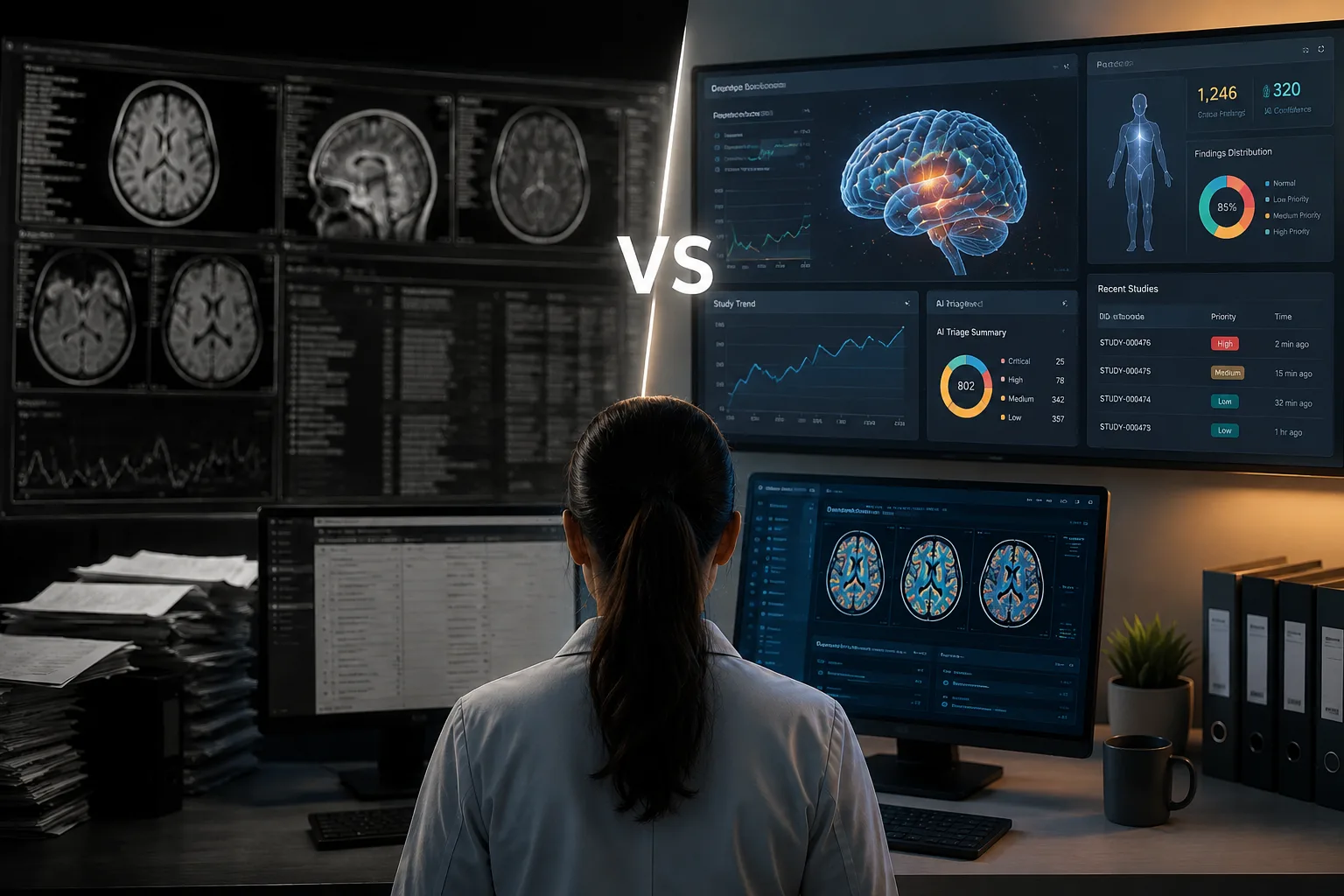
The conversation around AI in clinical imaging has reached an inflection point. While early adopters focused on automating isolated tasks — flagging image quality issues, measuring tumors, generating reports — the real transformation is happening at a different level entirely. The question is no longer whether AI can handle specific imaging tasks, but whether it can fundamentally redesign how clinical imaging operations function within regulated research.
This is the distinction between automation and intelligence. Automation replaces manual steps. Intelligence redesigns the entire workflow around better decision-making, predictive capability, and adaptive execution. For sponsors and CROs managing imaging endpoints in 2026, understanding this difference is critical to competitive advantage.
What Is AI in Clinical Imaging?
AI in clinical imaging encompasses machine learning models, computer vision algorithms, and intelligent orchestration systems designed to support medical image acquisition, analysis, quality assurance, and endpoint assessment in clinical trials. Unlike AI tools built for diagnostic radiology in clinical care, these systems operate under the regulatory constraints of Good Clinical Practice (GCP), 21 CFR Part 11 compliance, and protocol-specific acceptance criteria that vary across trials.
The technology stack typically includes:
- Computer vision models trained to detect anatomical structures, identify imaging artefacts, verify protocol compliance, and flag quality issues
- Natural language processing for extracting structured data from radiology reports and clinical narratives
- Workflow orchestration engines that route cases, manage reader assignments, trigger adjudication, and maintain audit trails
- Predictive analytics that forecast enrolment feasibility, identify sites at risk for protocol deviations, and optimize resource allocation
Critically, these systems are designed to augment, not replace, credentialed radiologists and clinical operations teams. The goal is not autonomous decision-making but rather intelligent assistance that allows human experts to operate at higher velocity and greater consistency.
The Evolution of AI in Clinical Research Imaging
The application of AI to clinical imaging has progressed through three distinct phases, each building on the limitations of the previous generation.
Phase 1: Task-Specific Automation (2015–2020)
Early implementations focused on narrow, well-defined tasks: automated lung nodule detection, tumor segmentation, and bone age assessment. These tools operated in isolation, requiring manual handoffs before and after the AI step. They delivered value in specific use cases but did not integrate into broader trial workflows. Radiologists treated them as decision support tools — helpful but not transformative.
The limitation of this phase was fragmentation. Each AI tool solved one problem but created integration challenges. Data had to be manually moved between systems. Audit trails were disconnected. And the overall workflow remained as manual as before, with AI functioning as an optional add-on rather than a core operational capability.
Phase 2: Workflow Integration (2020–2024)
The second wave brought AI into the operational layer. Systems began handling case routing, quality control checks, and structured data capture. Instead of standalone algorithms, AI became embedded in imaging platforms that connected to PACS, EDC systems, and project management tools. The focus shifted from “Can AI detect X?” to “Can AI make the entire imaging operation more efficient?”
This phase delivered measurable operational gains — faster turnaround times, fewer manual errors, better resource utilization — but remained largely reactive. AI responded to cases as they arrived rather than anticipating problems or optimizing across the trial lifecycle. The workflow was still predefined; AI simply executed it more efficiently.
Phase 3: Intelligent Orchestration (2024–Present)
The current generation treats AI as the orchestration layer for the entire imaging program. Rather than automating tasks within a fixed workflow, the system continuously optimizes the workflow itself based on real-time performance data, protocol requirements, and resource constraints. This means:
- Predictive site selection based on historical imaging protocol feasibility
- Adaptive quality thresholds that tighten or relax based on downstream read requirements
- Dynamic reader assignment that balances workload, expertise, and turnaround targets in real time
- Proactive deviation prevention through early warning systems that flag sites before problems compound
This is the shift from automation to intelligence. The platform doesn’t just execute predefined steps faster — it learns, adapts, and optimizes continuously. This is what platforms like ONIX AI™ deliver: not just faster execution of manual processes, but fundamentally redesigned workflows that were impossible without intelligent orchestration.
The Technical Architecture of AI-Native Imaging Platforms
Understanding how AI-native platforms differ from legacy systems requires looking at the underlying architecture. The distinction is not about whether AI is present, but where it sits in the technology stack.
Legacy Architecture: AI as a Feature
Traditional imaging CROs built their systems as service delivery platforms: PACS for storage, spreadsheets for tracking, email for communication, separate portals for reporting. When AI arrived, it was added as a feature — an image quality checker here, a measurement tool there — without changing the core architecture.
The result is a patchwork: AI outputs must be manually reviewed, transcribed, and moved between systems. The audit trail is fragmented. The workflow remains rigid. And when a protocol requires customization, it requires engineering work rather than configuration.
This approach delivers incremental improvements but leaves fundamental inefficiencies intact. The operational model remains manual-first, with AI providing assistance at specific touch points rather than driving the entire workflow.
AI-Native Architecture: Intelligence as the Foundation
AI-native platforms invert this model. The workflow engine itself is driven by machine learning models that continuously evaluate case characteristics, reader performance, protocol requirements, and operational constraints to make routing, prioritization, and quality decisions. Key architectural differences include:
- Protocol as Configuration: Instead of hardcoding workflows, the protocol document becomes a machine-readable configuration file that defines acceptance criteria, read requirements, adjudication rules, and data structures. Changes to the protocol update the system behaviour automatically without custom development.
- Event-Driven Orchestration: Every action — image upload, QC check, reader assignment, report submission — triggers the next step automatically based on protocol logic and current system state. There are no manual handoffs or batch processes waiting for human intervention.
- Unified Data Model: All imaging data, metadata, findings, and audit events are stored in a single relational database with full version control. This eliminates the fragmentation that creates compliance risk and enables real-time analytics across the entire imaging program.
- API-First Integration: The platform exposes all functionality through APIs, enabling seamless integration with EDC systems, CTMS platforms, and sponsor data warehouses. Structured data flows automatically rather than requiring manual export and import cycles.
This architecture enables the platform to scale across hundreds of concurrent trials without proportional increases in operational overhead — a fundamental economic advantage over legacy service models. When GenPhase AI’s imaging services deliver 30–40% faster read turnaround and 25%+ efficiency gains, it’s this architectural foundation that makes it possible.
Regulatory Considerations and Validation Requirements
AI tools used in clinical trials operate in a regulated environment. While they do not require FDA approval in the same way that diagnostic AI devices do, they must be validated for their intended use and documented appropriately in regulatory submissions.
Key Validation Requirements
- Algorithm Performance Documentation: Training data provenance, model architecture, performance metrics (sensitivity, specificity, accuracy) on independent validation sets, and edge case handling. Sponsors need to understand how the AI model was developed, what data it was trained on, and how it performs across different imaging modalities and patient populations.
- Version Control and Change Management: Procedures for tracking model versions, documenting changes, and revalidating when algorithms are updated. This is critical for maintaining consistency across the trial lifecycle. If an AI model is updated mid-trial, there must be a clear audit trail showing what changed, why, and how the new version was validated.
- Audit Trail Completeness: Demonstration that every AI-generated output (QC flag, case assignment, structured data field) is logged with timestamp, attribution, and traceability to source data. This is where 21 CFR Part 11 compliance becomes non-negotiable. Every decision the AI makes must be traceable, and every action must be attributable to a specific user or system process.
- Human Oversight Mechanisms: Documentation of how human reviewers validate, override, or approve AI-generated outputs before they become part of the trial record. This is the foundation of human-in-the-loop governance, ensuring that AI augments rather than replaces clinical judgment.
Practical Implications for Trial Sponsors
Sponsors should expect imaging partners to provide validation documentation as part of trial setup and to maintain validated, version-controlled AI models that can be audited during regulatory inspections. This documentation should include:
- Model validation reports with performance metrics
- Standard operating procedures for AI model deployment and monitoring
- Change control procedures for model updates
- Training records for staff using AI-assisted workflows
- Audit trail examples demonstrating full traceability
The regulatory landscape for AI in clinical trials is evolving, but the core principle remains constant: technology must enhance, not obscure, the integrity and traceability of clinical data.
Conclusion: Understanding the New Landscape
AI in clinical imaging has moved beyond the experimental phase. The technology exists, the regulatory framework supports it, and early adopters are demonstrating measurable operational advantages. But not all AI implementations are created equal.
The critical distinction is between AI as a feature and AI as infrastructure. Legacy imaging CROs are adding AI capabilities to existing manual workflows, delivering incremental improvements. AI-native platforms are rebuilding workflows from the ground up with intelligence as the foundation, delivering transformational change.
For sponsors and CROs evaluating imaging partners in 2026, the key questions are architectural:
- Is AI embedded in the workflow engine or bolted onto legacy systems?
- Does the platform learn and adapt, or simply execute faster?
- Is the audit trail unified and immutable, or fragmented across systems?
- Can protocols be configured rather than custom-coded?
The answers to these questions determine whether an imaging partner can deliver the speed, scale, and compliance that modern trials demand — or whether they’re simply rebranding legacy operations with AI marketing.
Ready to explore how AI-native platforms work in practice?
Learn more about ONIX AI™ or talk to our clinical team about your imaging program.
Explore ONIX AI™ Contact Our Clinical TeamFrequently Asked Questions
What is AI in clinical imaging?
AI in clinical imaging refers to machine learning algorithms and intelligent orchestration systems designed to support image acquisition, quality control, interpretation, and endpoint management in clinical trials. These systems operate within GCP and 21 CFR Part 11 compliance frameworks and are designed to augment rather than replace credentialed radiologists.
How is AI in clinical imaging different from diagnostic AI used in hospitals?
Diagnostic AI is designed for clinical care and focuses on detecting diseases or abnormalities to support patient treatment decisions. AI in clinical imaging is purpose-built for regulated research, operating under protocol-specific acceptance criteria and emphasizing audit trails, structured data capture, and workflow orchestration rather than autonomous diagnosis.
What are the three phases of AI evolution in clinical imaging?
Phase 1 (2015–2020) focused on task-specific automation like tumor segmentation. Phase 2 (2020–2024) integrated AI into workflows for case routing and QC. Phase 3 (2024–present) uses AI as the orchestration layer that continuously optimizes workflows based on real-time data and learns from outcomes.
What does “AI-native” mean in the context of clinical imaging platforms?
AI-native platforms are built from the ground up with AI as the core orchestration layer, where machine learning models drive workflow decisions, case routing, and quality control. This differs from “AI bolt-on” approaches where AI features are added to existing legacy systems without changing the underlying architecture.
Do AI tools used in clinical trials require FDA approval?
AI tools used in clinical trials do not require FDA approval in the same way that diagnostic AI devices do, but they must be validated for their intended use and documented appropriately in regulatory submissions. Validation documentation should include training data provenance, performance metrics, version control procedures, and audit trail completeness.
What is 21 CFR Part 11 compliance and why does it matter for AI in clinical imaging?
21 CFR Part 11 is the FDA regulation governing electronic records and electronic signatures in clinical trials. For AI in clinical imaging, this means every AI-generated output must be logged with timestamps, attribution, and traceability to source data in an immutable audit trail that can withstand regulatory inspection.
Can AI replace radiologists in clinical trials?
No. AI in clinical research is designed to augment, not replace, radiologists. Clinicians retain full accountability for medical interpretation and endpoint assessment. AI handles operational tasks — routing, QC flagging, data structuring — while radiologists focus on clinical judgment and protocol compliance.
What is “protocol as configuration” in AI-native platforms?
Protocol as configuration means the clinical trial protocol becomes a machine-readable file that automatically defines the system’s behaviour — acceptance criteria, read requirements, adjudication rules, and data structures. This eliminates custom coding for each trial and allows protocol changes to update system behaviour automatically.
How do I know if an imaging CRO is truly AI-native or just using AI marketing buzzwords?
Ask specific architectural questions: Is AI embedded in the workflow engine or added as a feature? Can protocols be configured or do they require custom development? Is there a unified audit trail or fragmented logs? Request demonstrations of intelligent case routing, pre-read QC, and structured data integration in action.
What should I look for in AI validation documentation from an imaging partner?
Look for model validation reports with performance metrics on independent datasets, standard operating procedures for AI deployment and monitoring, change control procedures for model updates, training records for staff, and audit trail examples demonstrating full traceability of AI-generated outputs.
Implementing AI in Clinical Imaging: Strategic Use Cases and Economic Impact
Above, we explored the conceptual foundation of AI in clinical imaging — the distinction between automation and intelligence, the evolution through three phases, and the architectural differences between AI-native and legacy platforms. Now we turn to the practical question that matters most to trial sponsors and CROs: where does AI deliver measurable impact, and how do you implement it without introducing execution risk?
The answer lies in understanding which operational bottlenecks create the most delay, cost, and compliance risk, and how intelligent orchestration addresses them. This isn’t about adopting technology for its own sake. It’s about solving specific, high-stakes problems that legacy manual processes cannot handle at the scale and velocity modern trials require.
Strategic Use Cases Where AI Delivers Measurable Impact
While AI’s potential in clinical imaging is broad, certain use cases deliver disproportionate strategic value. These are the areas where intelligent automation directly addresses high-cost pain points or unlocks capabilities that were previously infeasible.
Accelerating Study Start-Up
Traditional site activation for imaging trials takes 4–8 weeks: protocol training, imaging manual distribution, qualification scans, connectivity testing. Much of this is manual coordination across sites, vendors, and internal teams. Every email thread, every follow-up call, every manual check adds days to the timeline.
AI-native platforms compress this timeline to 72 hours by automating the entire activation sequence:
- Automated protocol configuration: The protocol document is parsed into machine-readable acceptance criteria, eliminating manual setup
- Site-specific imaging manual generation: The system automatically generates customized manuals based on site equipment and capabilities
- Interactive training modules: AI-driven comprehension checks ensure site staff understand protocol requirements before activation
- Remote connectivity validation: Automated test uploads verify PACS connectivity and image transfer protocols without manual coordination
For sponsors managing portfolios of trials, this acceleration compounds. A platform that activates sites in days rather than weeks can launch more trials in parallel, respond faster to competitive threats, and reduce the fixed costs of trial management infrastructure. When GenPhase AI targets 72-hour study activation timelines, it’s this end-to-end automation that makes it possible.
Enabling Real-Time Protocol Compliance Monitoring
Protocol deviations in imaging are expensive. A missed sequence, incorrect contrast timing, or wrong imaging plane can render a timepoint unusable — requiring a repeat scan, delaying dosing decisions, or creating missing data that complicates regulatory submissions.
Traditional QC catches these deviations after the read, when remediation is difficult or impossible. A radiologist spends time reviewing a non-compliant scan, discovers the problem, and initiates a query cycle that delays the trial and frustrates sites.
AI-powered pre-read QC inverts this model. Before any scan reaches a radiologist, the system evaluates it against protocol-defined acceptance criteria:
- Correct imaging modality (CT vs. MRI)
- Required sequences present (T1-weighted, T2-weighted, contrast-enhanced)
- Proper anatomical coverage (entire tumor visible, correct slice thickness)
- Acceptable image quality (no motion artefacts, appropriate contrast)
Cases that fail these checks are flagged immediately and routed back to the site for correction or clarification. Only protocol-compliant cases proceed to the read queue. This reduces post-read queries by 30–40% and ensures radiologists spend their time on interpretable, protocol-compliant imaging.
But the strategic value goes beyond individual case QC. Aggregated deviation data enables real-time site performance monitoring:
- Which sites have high deviation rates?
- Which specific protocol requirements are most frequently missed?
- Which imaging facilities consistently deliver compliant scans?
This intelligence allows proactive intervention — targeted retraining, site audits, or imaging vendor changes — before problems compound into data quality crises that jeopardize endpoints.
Optimizing Global Reader Networks
Managing a distributed network of subspecialty radiologists across time zones, languages, and workload capacities is operationally complex. Manual assignment by project managers introduces delays, creates bottlenecks when preferred readers are unavailable, and lacks the real-time adaptability needed to maintain SLA compliance.
AI-driven assignment systems treat this as a continuous optimization problem: match incoming cases to available readers based on:
- Subspecialty expertise: Neuro-radiologists for brain imaging, musculoskeletal specialists for orthopaedic trials
- Language requirements: Matching reader language capabilities with site locations
- Current workload: Balancing case volume across readers to prevent bottlenecks
- Historical turnaround performance: Prioritizing readers with consistent fast turnaround
- Time zone optimization: Routing cases to readers in active work hours for faster completion
The system learns from outcomes over time:
- Which readers excel at specific modalities?
- Which reader pairs have low adjudication rates (indicating consistent interpretation)?
- Which assignment patterns correlate with faster turnaround?
Over time, the network becomes more efficient without adding headcount. This is how ONIX AI™ delivers 24/7 global read coverage with 30–40% faster turnaround — not by hiring more radiologists, but by optimizing how existing capacity is utilized.
Supporting Adaptive Trial Designs
Adaptive trials require rapid endpoint assessment to inform dose escalation, cohort expansion, or futility decisions. Traditional imaging workflows — with manual routing, batch processing, and days-long turnaround — are incompatible with the operational tempo adaptive designs require.
AI-native platforms enable “fast-track” pathways for decision-critical time points:
- Priority routing: Cases flagged as decision-critical bypass the standard queue
- Real-time reader availability matching: The system identifies and assigns available readers immediately
- Automated adjudication triggering: Discrepancies are detected and routed to adjudicators without manual coordination
- Immediate structured data export: Results flow directly to decision-making systems via API
This capability transforms imaging from a lagging indicator (results available weeks after the scan) to a near-real-time input to trial decision logic — unlocking trial designs that were previously operationally infeasible.
The Economics of AI-Native vs. Legacy Imaging Operations
The business case for AI in clinical imaging is ultimately economic: does the technology reduce costs, compress timelines, or improve quality enough to justify the investment?
The answer depends on the scale and complexity of the imaging program. For small, simple trials with low case volumes, the operational overhead of legacy manual processes may be acceptable. But for large, multi-site, multi-modality trials with complex endpoints, the economics favour AI-native platforms decisively.
Cost Analysis: Phase III Oncology Trial Example
Consider a Phase III oncology trial with 300 sites, 1,000 patients, and serial imaging every 8 weeks over 18 months. Total imaging volume exceeds 6,000 time points. Managing this manually requires:
- Project managers to coordinate site activation and ongoing communication
- QC reviewers to check every scan post-read for protocol compliance
- Data coordinators to transcribe PDF reports into eCRF systems
- Operations leads to manually assign cases and manage reader workload
- Clinical teams to manage adjudication when readers disagree
An AI-native platform automates 60–70% of these tasks, reducing the operational team size by 40–50% while delivering faster turnaround and higher consistency. At scale, this translates to:
Both cost savings and timeline compression directly impact trial ROI and competitive positioning. In therapeutic areas where being first to market determines market share, weeks of timeline advantage can translate to hundreds of millions in revenue.
Scalability Economics
Equally important, AI-native platforms scale sub-linearly. Doubling case volume does not double operational costs because the intelligent orchestration layer handles increased complexity without proportional increases in human oversight.
Legacy models scale linearly: more cases require more project managers, more QC reviewers, more data coordinators. AI-native models scale logarithmically: the same platform and operational team can handle 2x, 5x, or 10x case volume with only incremental increases in infrastructure costs.
This economic advantage compounds over time. Organizations running multiple trials simultaneously can leverage a single AI-native platform across their entire portfolio, amortizing the technology investment across hundreds of studies.
Implementation Roadmap: How to Transition to AI-Native Imaging
For organizations currently using legacy imaging CROs, transitioning to an AI-native model requires careful planning. The goal is to capture operational benefits without introducing execution risk mid-trial.
Step 1: Assess Current State and Pain Points (Weeks 1–2)
Conduct a structured assessment of current imaging operations:
- Average site activation timeline
- Read turnaround times by modality and complexity
- Query volumes and resolution times
- Protocol deviation rates by site
- Operational team size and cost per read
Identify the highest-impact pain points — the areas where manual processes create the most delay, cost, or risk. These become the primary success metrics for the pilot.
Step 2: Pilot with a Non-Critical Trial (Months 1–3)
Select a Phase I or Phase II trial with moderate imaging complexity to pilot an AI-native platform. Avoid starting with a registration-enabling Phase III trial where execution risk is highest. The pilot objectives are:
- Validate that the platform can handle your specific protocol requirements
- Confirm performance improvements (site activation speed, read turnaround, query reduction)
- Train your team on the new system
- Build confidence before transitioning critical programs
Step 3: Validate Operational Metrics (Month 3)
Compare pilot performance against baseline metrics:
- Did site activation compress from weeks to days?
- Did read turnaround improve by the claimed 30–40%?
- Did query volumes decrease due to pre-read QC?
- Did structured data integration reduce eCRF transcription time?
Use this data to build the business case for broader adoption. Quantify the cost savings, timeline compression, and quality improvements in terms that resonate with executive stakeholders.
Step 4: Expand to Portfolio (Months 4–12)
Roll out the AI-native platform across the trial portfolio, starting with new studies and transitioning existing studies at natural inflection points: new cohort activations, protocol amendments requiring workflow changes, or site expansions into new geographic regions. Avoid forcing mid-trial transitions unless there’s a compelling operational reason. The goal is smooth adoption, not disruption.
Step 5: Optimize and Scale (Ongoing)
Continuously refine protocol templates, quality thresholds, and reader network configuration based on performance data. As the platform learns from more trials, operational efficiency compounds. Establish feedback loops between clinical operations teams and the technology platform to identify new automation opportunities and refine existing workflows.
The Competitive Landscape: Who’s Leading and Who’s Lagging
The clinical imaging CRO market is bifurcating. A small number of technology-forward providers are investing heavily in AI-native platforms, while the majority of legacy iCROs are adding AI features incrementally without rethinking their core architecture.
This creates a widening operational gap. AI-native providers can activate sites in days, deliver reads in hours, and scale across hundreds of concurrent trials with lean operational teams. Legacy providers remain constrained by manual processes, high fixed costs, and limited scalability.
Due Diligence Questions for Imaging Partners
- Is AI embedded in the workflow engine or bolted onto legacy systems? Ask to see the architecture diagram and understand where AI sits in the technology stack.
- Can the platform activate a site in 72 hours, or does it require weeks? Request case studies with documented activation timelines.
- Are reports structured and API-integrated, or PDFs requiring manual transcription? Ask for demonstrations of eCRF integration and structured data export.
- Is the audit trail unified and immutable, or fragmented across systems? Request audit trail documentation showing full traceability from image upload to final data export.
- Can the vendor provide validation documentation for AI models? Ask for model validation reports, performance metrics, and change control procedures.
The answers reveal whether a provider is genuinely AI-native or simply rebranding legacy operations with AI marketing.
Conclusion: Intelligence as Infrastructure
AI in clinical imaging is no longer an experimental technology — it is rapidly becoming table stakes for competitive trial execution. The organizations that recognize this shift early and build their imaging operations around intelligent orchestration will capture significant advantages in speed, cost, and quality.
But this transition requires more than adopting new tools. It requires rethinking how imaging operations are structured, how teams are organized, and how technology and clinical expertise combine to deliver regulatory-grade execution at scale.
The future of clinical imaging is not radiologists replaced by algorithms. It is credentialed clinicians empowered by intelligent systems that handle operational complexity, enabling them to focus on medical judgment, protocol adherence, and patient safety.
For sponsors and CROs ready to make this transition, the path is clear: evaluate your current imaging operations, identify the highest-impact pain points, pilot an AI-native platform, and scale based on validated performance improvements.
Ready to implement AI-native imaging execution?
Explore ONIX AI™ or talk to our clinical team about your imaging challenges and implementation roadmap.
Explore ONIX AI™ Contact Our Clinical TeamFrequently Asked Questions — Part Two
What are the most impactful use cases for AI in clinical imaging?
The highest-impact use cases are accelerating study start-up (compressing 4–8 weeks to 72 hours), enabling real-time protocol compliance monitoring through pre-read QC, optimizing global reader networks for 30–40% faster turnaround, and supporting adaptive trial designs with near-real-time endpoint assessment.
How much does AI-native imaging reduce operational costs?
For large Phase III trials with 6,000+ imaging timepoints, AI-native platforms can reduce operational team size by 40–50% and deliver $2–3 million in cost savings over the trial lifecycle through automation of case routing, QC, data transcription, and adjudication management.
How long does it take to implement an AI-native imaging platform?
A phased implementation typically takes 3–12 months: 1–2 weeks for assessment, 1–3 months for a pilot with a non-critical trial, 1 month for metrics validation, and 4–12 months for portfolio-wide rollout. Individual study activation on an established platform can occur in 72 hours.
What should I pilot first when transitioning to AI-native imaging?
Start with a Phase I or Phase II trial with moderate imaging complexity — enough to test the platform’s capabilities but not so critical that execution risk threatens regulatory timelines. Avoid starting with registration-enabling Phase III trials until the platform is validated.
How do I measure success during an AI imaging pilot?
Compare baseline metrics to pilot performance: site activation timeline (weeks to days), read turnaround time (30–40% improvement target), query volumes (reduction due to pre-read QC), and eCRF transcription time (elimination through structured data integration).
Can AI-native platforms handle complex, multi-modality trials?
Yes. AI-native platforms are specifically designed for protocol complexity. The “protocol as configuration” approach means acceptance criteria, read requirements, and adjudication rules are defined per protocol without custom coding, making complex multi-modality trials easier to manage than on legacy systems.
What’s the difference between AI-native and legacy imaging CROs adding AI features?
AI-native platforms are built from the ground up with AI driving workflow orchestration, case routing, and quality decisions. Legacy CROs add AI features (image quality checkers, measurement tools) to existing manual workflows without changing the underlying architecture, delivering incremental rather than transformational improvements.
How does AI in clinical imaging support adaptive trial designs?
AI enables fast-track pathways for decision-critical time points through priority routing, real-time reader availability matching, automated adjudication triggering, and immediate structured data export via API — transforming imaging from a weeks-long lagging indicator to a near-real-time decision input.
What due diligence should I conduct when evaluating AI imaging partners?
Ask architectural questions: Is AI embedded in the workflow engine? Can sites be activated in 72 hours? Are reports structured and API-integrated? Is the audit trail unified? Request demonstrations of pre-read QC, intelligent routing, and structured data integration, plus validation documentation for AI models.
How do AI-native platforms scale compared to legacy operations?
AI-native platforms scale sub-linearly — doubling case volume doesn’t double operational costs because intelligent orchestration handles complexity without proportional increases in human oversight. Legacy operations scale linearly, requiring more staff for more cases, making AI-native platforms increasingly cost-advantageous at scale.